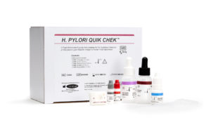
Two new FDA-cleared immunoassays added to test choices for non-invasive diagnosis of gastritis associated with H. pylori

Introduction

The 2017 ACG Guidelines recommend non-invasive tests which detect current disease to diagnosis H. pylori infection in patients under the age of 55 who are not exhibiting alarm symptoms (1). Non-invasive H. pylori testing options include stool antigen testing, urea breath testing, and serology (1, 2). Laboratory directors must decide how to optimally combine test accuracy, cost, ease-of-use and time-to-result. Two new FDA-cleared immunoassays, the first in 15 years, are now available. The strong performance of these new bench-top rapid stool antigen tests and their sample flexibility offer several advantages over alternative testing methods.


H. pylori microbiology influences test design and choice

H. pylori bacteria are specialized for growth in the harsh acidic environment of the stomach (3, 4). The bacteria use flagella to propel the organism into and under the mucus layer that normally protects the epithelial cells of the stomach. Although this mucus layer is much less acidic that the lumen, H. pylori further adjusts the pH of its mucus niche. H. pylori expresses a urease enzyme that breaks down urea into ammonia to increase local pH while releasing carbon dioxide. This reaction is uncommon in other stomach bacteria An illustration of H. Pyloriand is the basis of some testing methods (4).

Unlike many other gastrointestinal bacteria, H. pylori typically is not cultured from fecal specimens or even from endoscopic biopsy samples unless antibiotic resistance testing is required. However, antibiotic resistance is an increasing problem (5, 6). Eradication of H. pylori can no longer be assumed after standard antibiotic therapy. This has led to adoption of a Test- Treat-Test strategy in which patients undergo follow-up testing four weeks following eradication therapy to determine if the bacteria have been successfully eliminated (1, 5, 7). A non-invasive stool antigen test or urea breath test (UBT) is favored for this follow-up testing to avoid the expense and discomfort of endoscopy and biopsy for the patient. Serology that detects antibodies to H. pylori cannot be used for follow-up, as antibodies persist even after successful eradication (1).


Patients can also influence test results

H. pylori infections and associated symptoms and inflammation can wax and wane. Patients who seek medical care for symptoms are often experiencing active inflammation and may already be taking a proton pump inhibitor (PPI) or bismuth-containing antacid. These compounds can alter the reliability of both UBTs and stool antigen tests by decreasing inflammation (1).

For this reason, UBTs require discontinuation of supportive therapies. With stool antigen testing, a positive result is reliable, a negative result in a patient still suspected of having an H. pylori infection may warrant discontinuation of supportive therapy and retesting. Not surprisingly, patients dislike stopping these supportive therapies for 10-14 days prior to testing. Compliance rates are unknown but circumstantial evidence suggests that testing laboratories may wish to request confirmation of patient PPI status.

Also, an unknown fraction of patients may have current or recent antibiotics exposure. Antibiotic treatment within four weeks prior to testing may suppress bacteria below detectable levels but not eradicate them fully. Thus, antibiotics therapy should be completed prior to these tests to avoid false negative results (1).

Table 1. Test performance of non-invasive H. pylori assays

TestSensitivity
Specificity
Diagnose Current Disease?Confirm Eradication?
Serology for H. pylori Antibody80 - 84%
<80%
NoNo
Urea Breath Test (UBT)97%
95%
YesYes
Stool Antigen
96 wellH. PYLORI CHEK1100%
96%
YesYes
Premier Platinum HpSA® PLUS 296%
96%
YesYes
RapidH. PYLORI QUIK CHEK197%
100%
YesYes
ImmunoCard STAT!® HpSA® 291%
92%
YesYes

1. H. PYLORI CHEK and H. PYLORI QUIK CHEK Package Inserts.
2. Premier Platinum HpSA® PLUS and ImmunoCard STAT!® HpSA® Package Inserts.


Basis for test choices

Serology

Serology is a widely used test for H. pylori infection. The technology is simple, non-invasive, inexpensive and can be performed rapidly with a specimen that is easy to collect. Unfortunately, in many areas of the world over half of the population has been infected with H. pylori (3). In the United States, clinical practices that include immigrant or disadvantaged populations can find that 60-80% of these individuals have H. pylori antibodies. In these locations, serum tests for H. pylori antibodies are not useful as patients with active H. pylori gastritis cannot be discriminated from others that simply carry H. pylori antibodies from previous infection (1, 4). Overall, serology tests have lower sensitivity and specificity than other test types (Table 1). As a result, an increasing number of providers of health care insurance no longer reimburse for H. pylori serology testing (8). Some laboratories and health care providers are also discontinuing H. pylori antibody tests. For example, Quest and the Mayo Clinic have not offered this test since 2016. Due to guideline recommendations and inferior test performance, several insurers including Cigna, Aetna, Anthem Blue Cross Blue Shield, and Geisinger Health Plan have stopped reimbursing H. pylori serology testing.

Urea breath tests

UBTs take advantage of the urease activity of H. pylori. To perform the test, patients exhale into a sample collection device before and after drinking a liquid containing either 13C- or 14C-labeled urea. The timing of sample collection after ingestion of the labeled drink, typically 15- 30 minutes, allows H. pylori metabolism of the labeled urea, absorption of the labeled CO2 into the blood stream and its release into the lungs. A major drawback of UBTs is the cost. The instrument and software to measure and analyze the labeled CO2 are expensive. Isotope ratio mass spectrometers (IRMS) used for measurement of 13C enrichment are costly. Isotope-selective infrared spectrometers are a new, less expensive alternative. 13C-urea is stable but is relatively high-priced; 14C-labeled urea is inexpensive but radioactive. Samples derived from 14C-labeled urea require the facility to be licensed for use and disposal of radioactive agents. These costs are absorbed by the laboratory or are passed on to patients. While sample collection is non-invasive, it is time-consuming and ingestion of radioactive material, particularly for children and pregnant women, is undesirable. Nonetheless, UBTs have good sensitivity and specificity, test for current disease, and can detect H. pylori that is present in any part of the stomach or that is dispersed in patches (Table 1) (5).

Table 2. Initial diagnosis H. PYLORI QUIK CHEK™ and H. PYLORI CHEK™ tests versus Composite Reference Method (CRM)

N=122CRM PositiveCRM Negative
H. PYLORI QUIK CHEK™ Positive320
H. PYLORI QUIK CHEK™ Negative1*89
95% Confidence Limits
Sensitivity97.0%84.7% - 99.5%
Specificity100.0%95.9% - 100.0%

*Additional testing with an FDA-cleared H. pylori stool antigen test provided an antigen-negative result.

N=109CRM PositiveCRM Negative
H. PYLORI CHEK™ Positive323*
H. PYLORI CHEK™ Negative074
95% Confidence Limits
Sensitivity100.0%89.3% - 98.9%
Specificity96.1%89.2% - 98.7%

*All three specimens tested positive initially in the H. PYLORI CHEK test, but negative upon re-testing with the H. PYLORI CHEK test.


Stool antigen tests

Stool antigen tests are simple to perform, involve a sample that can be collected non-invasively, can be batched, and are less expensive than UBTs. Stool antigen tests are specifically designed to identify current infection by detecting antigen(s) that are produced by live H. pylori bacteria and shed into the stool. Stool tests also detect infection from throughout the stomach and duodenum. Importantly, stool antigen tests are more accurate than serology for detecting current, active infections. Because of these characteristics, stool tests are a good first choice for initial detection of H. pylori, for confirming success of treatment or eradication therapy, as well as for patients that may need frequent monitoring such as H. pylori-infected pregnant women (5).

The first new stool antigen tests in 15 years, the H. PYLORI QUIK CHEK™ and the H. PYLORI CHEK™ tests were FDA-cleared in 2018. Similar to the commercially available PREMIER and ImmunoCard STAT! HpSA assays, these new tests utilize immobilized H. pylori-specific antibodies to capture an antigen and antibody conjugates to detect the immunocomplex.

The performance of the H. PYLORI QUIK CHEK™ test and the H. PYLORI CHEK™ tests were evaluated at five independent sites. Patients who were undergoing endoscopy as part of routine care were recruited for the study. No difference in test performance was observed based on patient age or gender. The H. PYLORI CHEK™ and H. PYLORI QUIK CHEK™ tests exhibit excellent sensitivity and specificity that was verified against a Composite Reference Method (CRM) of histology and rapid urease testing performed on biopsies collected during endoscopy (Table 2). The H. PYLORI QUIK CHEK™ test exhibited a sensitivity of 97.0% and specificity of 100.0% with CRM biopsy results. The H. PYLORI CHEK™ test exhibited sensitivity of 100.0.% and specificity of 96.1% with CRM biopsy results.

Table 3. Retrospective sample study

N=200Commercial ELISA PositiveCommercial ELISA Negative
H. PYLORI QUIK CHEK™ Positive933*
H. PYLORI QUIK CHEK™ Negative1**103
95% Confidence Limits
Percent Positive Agreement98.9%94.2% - 99.8%
Percent Negative Agreement97.2%92.0% - 99.0%

*H. pylori DNA was amplified from the samples with PCR

**No H. pylori DNA was amplified from the samples with PCR

N=196Commercial ELISA PositiveCommercial ELISA Negative
H. PYLORI CHEK™ Positive750
H. PYLORI CHEK™ Negative0121
95% Confidence Limits
Percent Positive Agreement100.0%95.1% - 100.0%
Percent Negative Agreement100.0%96.9% - 100.0%

In a supplemental study of retrospective samples, the H. PYLORI QUIK CHEK™ and H. PYLORI CHEK™ tests were compared to another FDA-cleared commercial ELISA. For this study, 200 samples (96 positive and 104 negative) were tested by the H. PYLORI QUIK CHEK™ test and 196 samples (75 positive and 121 negative) were evaluated with the H. PYLORI CHEK™ test (Table 3). There are no FDA-cleared PCR tests for H. pylori. This may be due, in part, to DNA degradation in either biopsy or fecal samples. The antigen detected in the H. PYLORI CHEK™ and H. PYLORI QUIK CHEK™ tests is highly stable.

The H. PYLORI QUIK CHEK™ test detects H. pylori in 30 minutes, while the H. PYLORI CHEK™ test can be performed with or without automation in one hour. The kits also offer flexible specimen transport and storage conditions including room temperature storage of specimens and the use of C&S and Cary Blair transport media (Table 4). No conversion of positive-to-negative or negative-to-positive samples was observed when comparing fresh to frozen samples at three time points over two weeks. The adaptable specimen transport conditions were tested rigorously to simplify sample collection and improve compliance by patients and laboratory confidence in sample results. The analytical sensitivity of both tests were similar in fresh samples or in transport media.

The H. PYLORI CHEK™ and H. PYLORI QUIK CHEK™ tests demonstrate robust performance compared to serology, UBTs and currently marketed stool antigen tests. The H. PYLORI CHEK™ and H. PYLORI QUIK CHEK™ tests are accurate as a test of cure when utilized > 4 weeks after treatment.

Table 4. Sample types and storage time for H. PYLORI CHEK™ and H. PYLORI QUIK CHEK™ tests

Sample TypeStorage ConditionRecommended Storage Time
Fresh Unpreserved, Cary Blair or C&S Transport Media2ºC - 8ºC96 hours
20ºC - 25ºC96 hours
Frozen Unpreserved Samples≤ -10ºC14 days


Summary

The H. PYLORI QUIK CHEK™ and H. PYLORI CHEK™ Stool Antigen Tests:

  • Are patient-friendly, economical, and convenient
  • Detect current infections
  • Show robust performance with real-life clinical specimens compared against histology and rapid-urease assays from biopsy
  • Are a good choice for initial diagnosis, for confirming treatment efficacy, and for patients that require frequent monitoring
  • Give accurate positive results for patients taking PPIs

References

  1. Chey WD, Leontiadis GI, Howden CW, Moss SF. 2017. ACG Clinical Guidelines: Treatment of Helicobacter pylori Am J Gastroenterol 112:212-238.doi: 10.1038.
  2. Talley NJ. 2005. American Gastroenterological Association Medical Position Statement: Evaluation of Dyspepsia. Gastroenterology 129:1753-1755.doi: 10.1053/j. gastro.2005.09.019.
  3. Burkitt MD, Duckworth CA, Williams JM, Pritchard DM. 2017. Helicobacter pylori-induced gastric pathology: insights from in vivo and ex vivo Disease Models & Mechanisms 10:89-104.doi: 10.1242/dmm.027649.
  4. Kusters JG, van Vliet AHM, Kuipers EJ. 2006. Pathogenesis of Helicobacter pylori Clinical Microbiology Reviews 19:449-490.doi: 10.1128/cmr.00054-05.
  5. Testerman TL, Morris J. 2014. Beyond the stomach: An updated view of Helicobacter pylori pathogenesis, diagnosis, and treatment. World J Gastroenterol 20:12781-12808.doi: 10.3748/wjg.v20.i36.12781.
  6. Park JY, Dunbar KB, Mitui M, Arnold CA, Lam- Himlin DM, Valasek MA, Thung I, Okwara C, Coss E, Cryer B, Doern CD. 2016. Helicobacter pylori Clarithromycin Resistance and Treatment Failure Are Common in the USA. Digestive Diseases and Sciences 61:2373-2380.doi: 10.1007/s10620-016-4091-8.
  7. Malfertheiner P, Megraud F, O’Morain CA, Atherton J, Axon ATR, Bazzoli F, Gensini GF, Gisbert JP, Graham DY, Rokkas T, El-Omar EM, Kuipers EJ. 2012. Management of Helicobacter pylori infection– the Maastricht IV/ Florence Consensus Report. Gut 61:646-664.doi: 10.1136/gutjnl-2012-302084.
  8. El-Serag HB, Kao JY, Kanwal F, Gilger M, LoVecchio F, Moss SF, Crowe S, Elfant A, Haas T, Hapke RJ, Graham DY. 2018. Houston Consensus Conference on Testing for Helicobacter pylori Infection in the United States. Clinical Gastroenterology and Hepatology 16:992-1002.e1006.10.1016/j. cgh.2018.03.013.